Tập tin:ClpB.gif

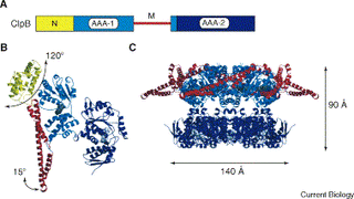
Figure 1. The crystal structure of T. thermophilus ClpB.(A) Domain organisation of Hsp104/ClpB. The two AAA domains, although highly conserved, exhibit significant differences and can be classified accordingly as AAA-1 and AAA-2. The middle (M) domain is specific for Hsp104/ClpB and serves for Hsp104/ClpB protein classification. (B) Structure of monomeric T. thermophilus ClpB. The colour code for the individual domains is the same as in (A). AMPPNP is shown in grey. Movements of N and M domains are indicated. (C) Hexameric model of ClpB in the AMPPNP-bound state, based on single-particle reconstitution using cryo-electron microscopy. Height and diameter of the ClpB oligomer is given. The mobile N domains are not visible in the single-particle reconstitution, presumably because they are flexible. M domains were only partially resolved and were placed according to their positioning in the crystal structure [11.].
Lịch sử tập tin
Nhấn vào một ngày/giờ để xem nội dung tập tin tại thời điểm đó.
| Ngày/Giờ | Hình nhỏ | Kích cỡ | Thành viên | Miêu tả | |
|---|---|---|---|---|---|
| hiện | 16:10, 4/8/2006 |  | 379×213 (17 kB) | Cao Xuân Hiếu (Thảo luận | đóng góp) | Figure 1. The crystal structure of T. thermophilus ClpB.(A) Domain organisation of Hsp104/ClpB. The two AAA domains, although highly conserved, exhibit significant differences and can be classified accordingly as AAA-1 and AAA-2. The middle (M) domain is |
- Bạn không có thể ghi đè lên tập tin này.
Các trang sử dụng tập tin
Trang sau có liên kết đến tập tin này:

