Chủ đề nóng: Phương pháp kỷ luật tích cực - Cổ học tinh hoa - Những thói hư tật xấu của người Việt - Công lý: Việc đúng nên làm - Giáo án Điện tử - Sách giáo khoa - Học tiếng Anh - Bài giảng trực tuyến - Món ăn bài thuốc - Chăm sóc bà bầu - Môi trường - Tiết kiệm điện - Nhi khoa - Ung thư - Tác hại của thuốc lá - Các kỹ thuật dạy học tích cực
- Dạy học phát triển năng lực - Chương trình giáo dục phổ thông
Tập tin:Buchnera.jpg
Từ VLOS

Kích thước của hình xem trước: 320×212 điểm ảnh. Độ phân giải khác: 803×533 điểm ảnh.
Tập tin gốc (803×533 điểm ảnh, kích thước tập tin: 202 kB, kiểu MIME: image/jpeg)
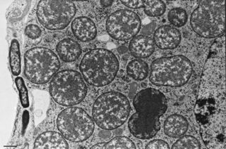
This electron micrograph from a pea aphid (Acyrthosiphon pisum) shows the obligate symbiont Buchnera aphidicola (large round cells in the center) within an aphid bacteriocyte, the aphid nucleus (far right), facultative symbionts (rod-shaped bacteria on the far left, within a separate aphid cell), and mitochondria (scattered among the Buchnera cells, within the bacteriocyte). Scale bar = 1 micron. Photo by J. White and N. Moran, University of Arizona.
http://www.current-biology.com/content/article/image?uid=PIIS0960982206022123&imageid=fig2
Lịch sử tập tin
Nhấn vào một ngày/giờ để xem nội dung tập tin tại thời điểm đó.
| Ngày/Giờ | Hình nhỏ | Kích cỡ | Thành viên | Miêu tả | |
|---|---|---|---|---|---|
| hiện | 16:48, 4/11/2006 | | 803×533 (202 kB) | Cao Xuân Hiếu (Thảo luận | đóng góp) | This electron micrograph from a pea aphid (Acyrthosiphon pisum) shows the obligate symbiont Buchnera aphidicola (large round cells in the center) within an aphid bacteriocyte, the aphid nucleus (far right), facultative symbionts (rod-shaped bacteria on th |
- Bạn không có thể ghi đè lên tập tin này.
Các trang sử dụng tập tin
Trang sau có liên kết đến tập tin này:

